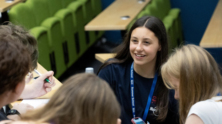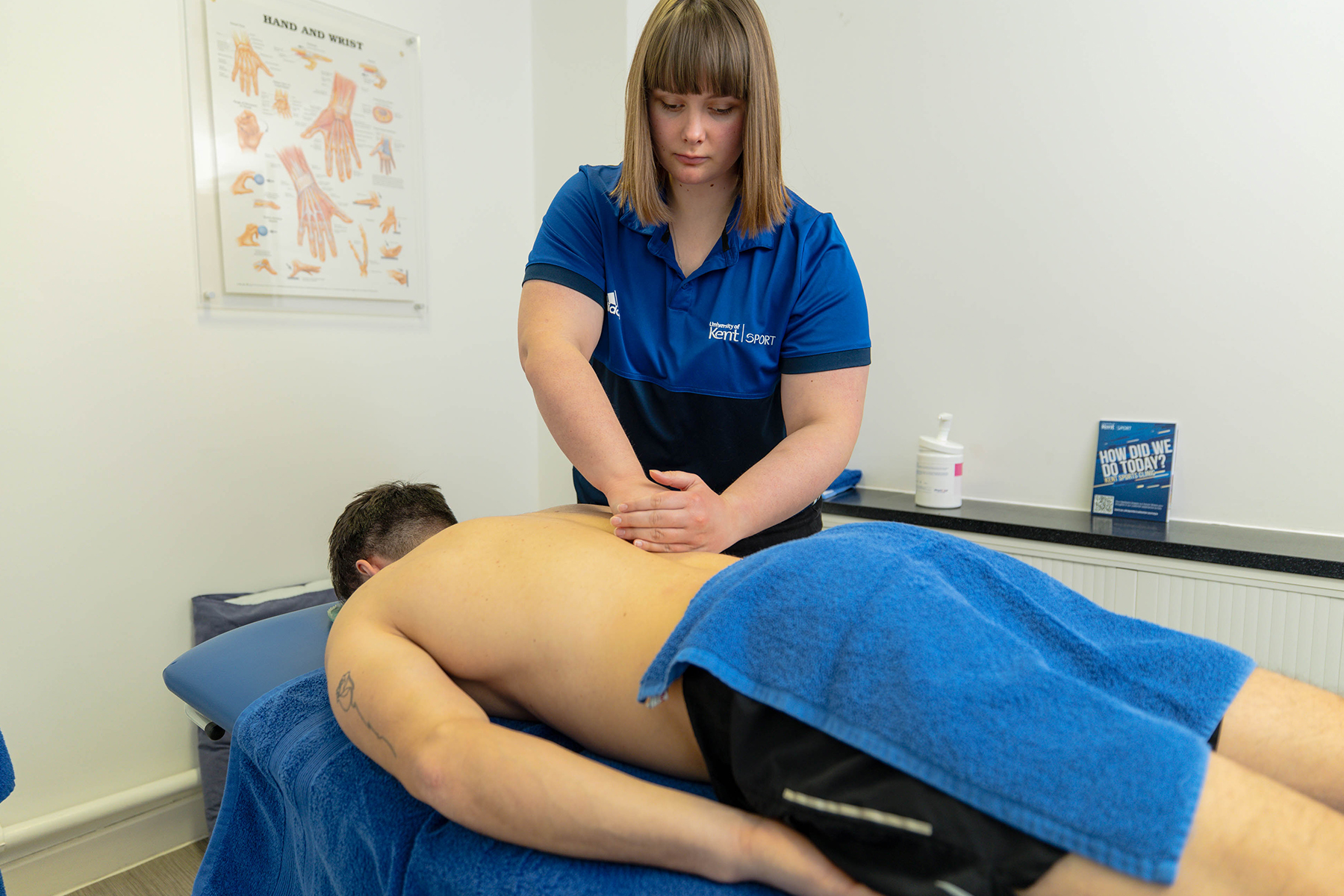
Every August, the Bioscience department, in the School of Natural Sciences, hosts a Bioscience Work Experience Week for students in year 12 and above. The role of this week is to give the students hands-on lab experience, learn about coming to university and discover what it is like to study a bioscience subject in higher education. Daisy Shaw, PhD in Microbiology and postgraduate helper, shares her experience of supporting this annual event.
‘The outreach week has been running since 2017, and was originally set up by Professor Ben Goult, Dr Rosalyn Masterton and Dr Anastasios Tsaousis. For the last two years Dr Rosalyn Masterton has organised it alongside Dr Emma Hargreaves and Dr Dave Beal. All five of these academics have worked incredibly hard to provide the students with an enriching experience, with support from Dr Katrine Solvaag, who co-ordinates student recruitment and engagement in advance.
‘Many students who attended past work experience weeks have gone on to enrol at Kent for their undergraduate degree, with some even continuing on to undertake a PhD. My fellow postgraduate demonstrator, Matt, is one of those ‘success stories’, having attended the first ever Bioscience Work Experience Week in 2017.
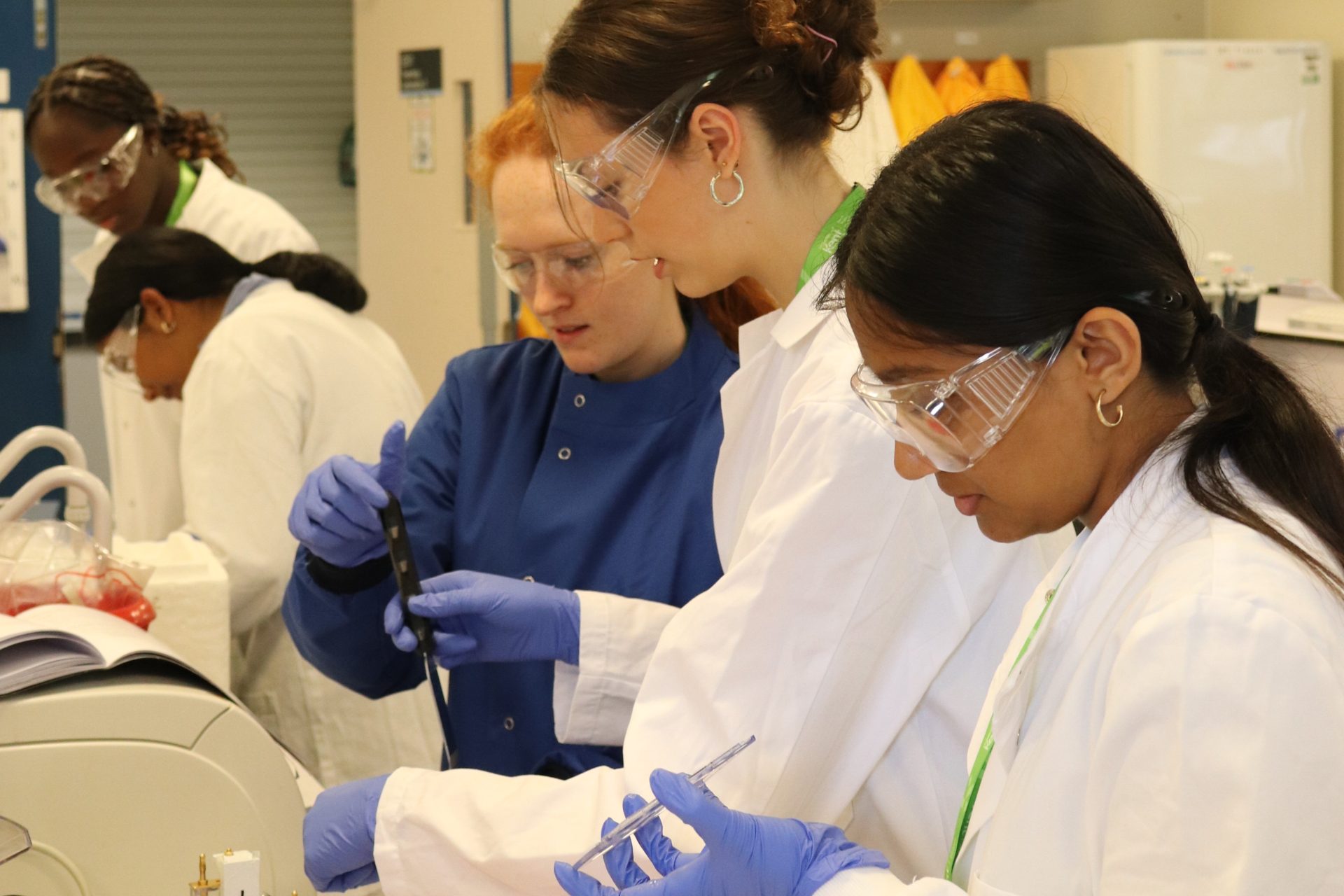
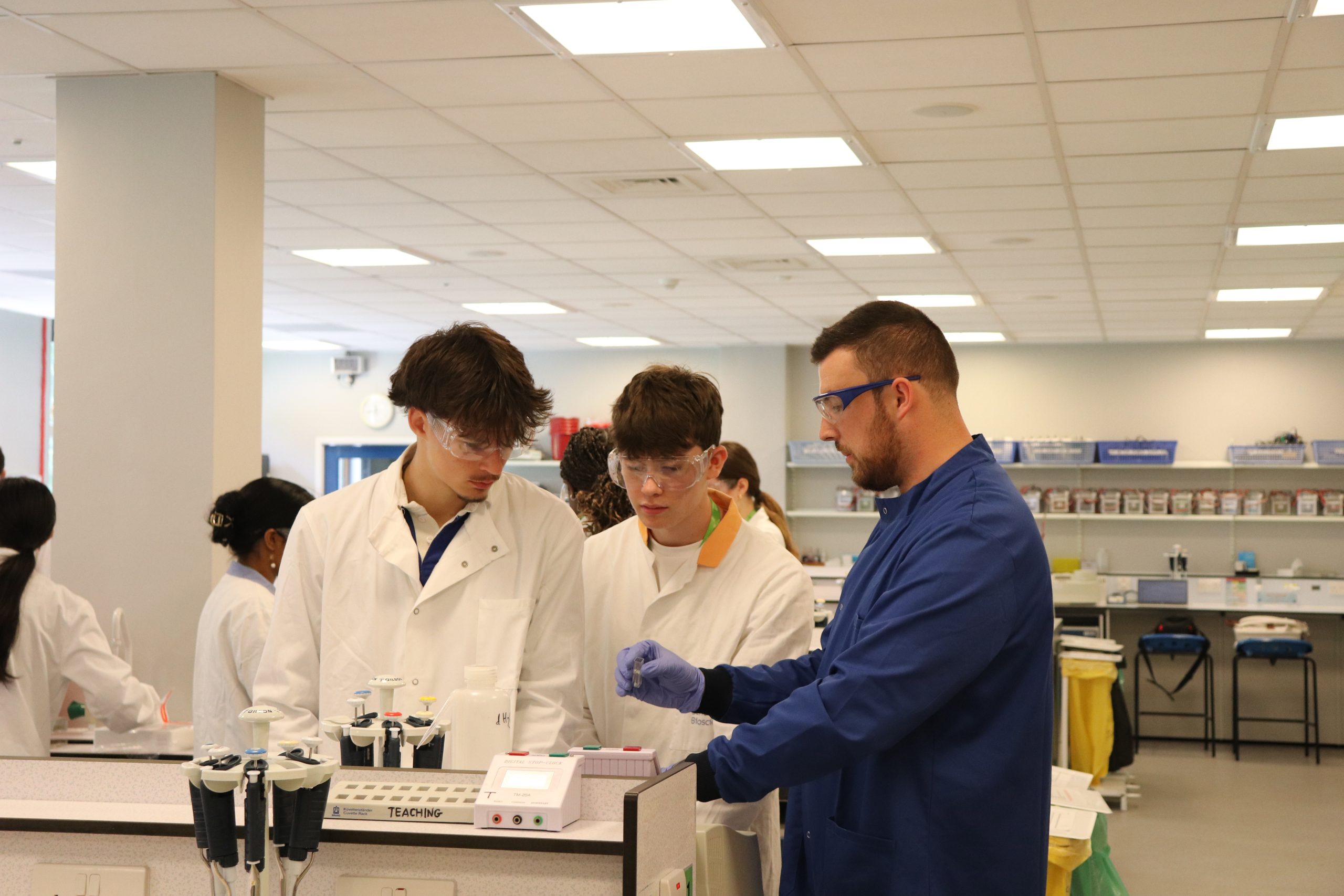
‘So, what did the students get up to? Well, they started the week with an introduction to their five-day project; to genetically engineer E. coli bacteria to express different fluorescent proteins, then extract, purify and characterise this protein. This technique is commonly used by scientists to visualise the movement of proteins in cells, understand how they work and identify new drug-delivery pathways which could lead to the development of new treatments for disease.’
PhD student Matthew Rice (in blue) took part in Bioscience Week when he was a school student and is now a co-founder at DrugUptech, a biotech company that is providing smarter compound uptake analysis to accelerate early-stage discovery in agritech and drug development.
Day 1: Setting up the experiment
‘On Monday the students learnt the basics of working in the lab, such as how to maintain a lab book, using a micropipette and working aseptically under a Bunsen burner. They also made bacterial growth media and set up agar plates, then introduced the DNA which produces fluorescent proteins into the E.Coli cells, before leaving them to grow overnight on the agar plates.’
Day 2: Troubleshooting the problem
‘The next day, the students learnt how to observe bacterial growth using a spectrophotometer which measures how much light is absorbed when passed through a sample. Up until this point, their E. coli was happily growing, but their protein of interest wasn’t being produced, so they added a special chemical called IPTG which allows the bacteria to start producing large amounts of their protein. Even just a few hours after induction, their fluorescent proteins were becoming visible! They left these to grow overnight, and the next morning were greeted with super colourful cultures.’
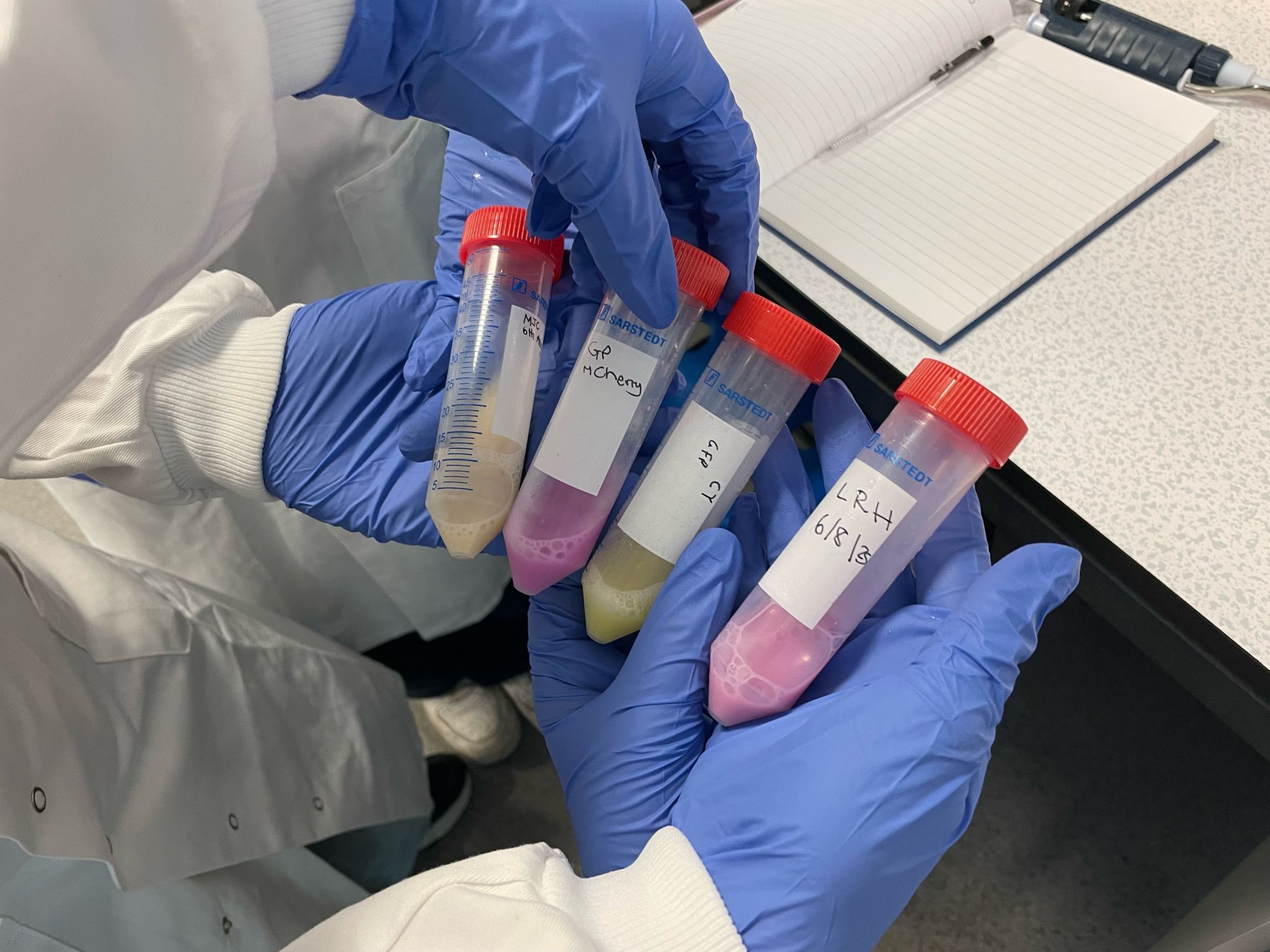
Students added a chemical called IPTG to their bacterial samples to make them express the fluorescent proteins which give them their colour.
Day 3: Extracting proteins
‘Their task on Wednesday was to extract and purify their proteins of interest. They did this by separating the bacterial cells from the media using a technique called centrifugation. The cells then underwent sonication, which breaks open (or ‘lyses’) the bacterial cells using high frequency sound waves, releasing all the proteins within them. To isolate the fluorescent proteins alone they used a technique called nickel ion affinity chromatography.’
Day 4: Sorting the proteins.
‘The following day, they separated proteins by size using polyacrylamide gel electrophoresis, producing an SDS-page gel and an immunoblot.’
Day 5: Sharing the outcomes of their experiment
‘On the final day, students learnt about one of the most important parts of science – dissemination! They spent the first part of the day producing posters of their methods and results, ready to show these to their parents in the afternoon.
‘They also had the chance to be creative with some agar art. The agar plates acted as their canvas, and the bacterial cultures containing the fluorescent proteins acted as their paint. The results were fantastic, with designs including jellyfish, flowers, stars, a turtle and even Walter White! A shortlist was made and the final 10 were voted on by the students and their parents in the closing ceremony. Students were also encouraged to take interesting pictures throughout the week in the lab, and a winner was selected from these too.
‘This was my fourth year helping with this event, and as a postgraduate helper, my role is to guide the students with their lab work, provide demonstrations and answer any questions they might have about the university or pursuing biosciences further. I get a lot out of this week every year, but most of all it is rewarding to see the students grow in confidence in the lab as the week goes on.’