We’re sorry for any disruption, but this is going to be a very exciting addition to the school.
If you can hear some drilling and things are a bit dusty at The Marlowe building it’s because the school is in the process of preparing the new Imaging Centre for Life Sciences.
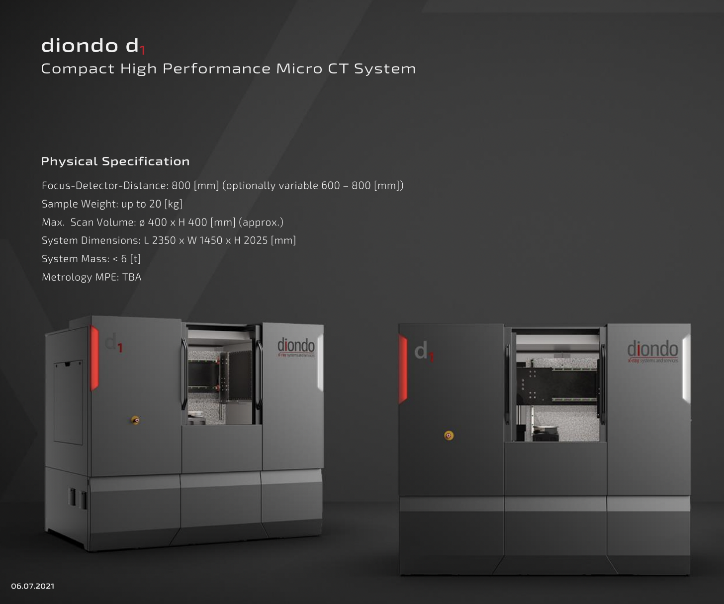
Housing a state-of-the-art microtomographic scanner that can produce 3D X-rays of objects at very high resolution, the Imaging Centre for Life Sciences will enable researchers from across the University of Kent, as well as external users, to examine the internal structures of a multitude of objects in incredible detail. Common applications include materials science, palaeontology, experimental biology and archaeology, and there are currently less than five in the UK.
Having digitised specimens means they are far more accessible for learning and research, and we will be able to teach students technical skills that are becoming increasingly important within Biological Anthropology and, Biological Sciences more broadly.
The new Imaging Centre has largely been the initiative of Professor Tracy Kivell and Professor Matthew Skinner. The Diondo D scanner ‘in my opinion, looks pretty exciting’ Matthew told us. ‘The machine weighs 5 tonnes! So we are having to prepare the floor and design a space around it.’

The whole department are really excited about what this means for historical discoveries as well as preparing ourselves for the future, for advances in research and in teaching.
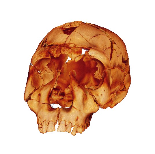
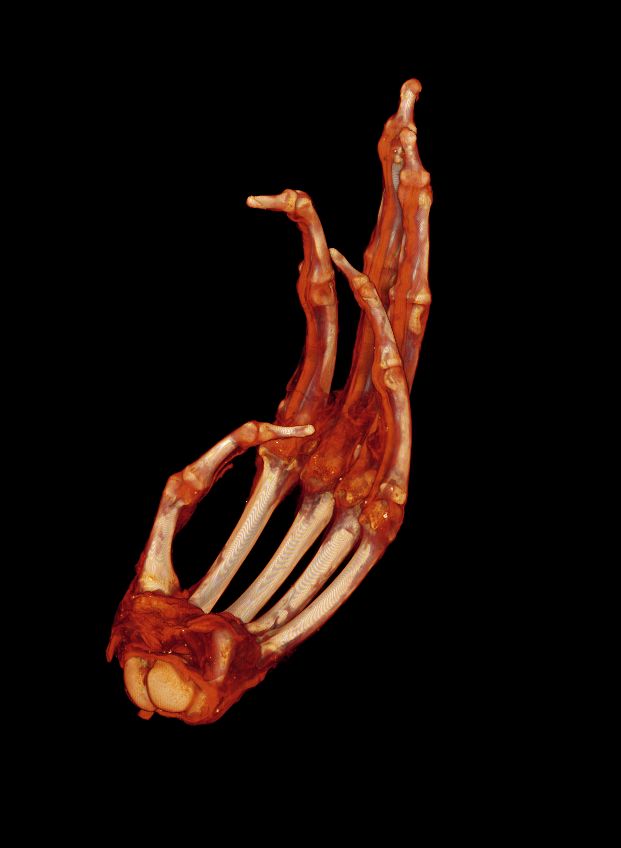
‘The machine is incredibly capable. The part I am most excited about (personally) is the resolution.’ Lecturer in Biological Anthropology Dr Devin Finaughty told us ‘As a micro-CT scanner, it can provide insanely detailed insight into the internal structure of whatever we put inside it. But the real fun is found with what we can do with that insight. Apart from being able to non-destructively visualise parts of artefacts, fossils, and other biological material that would otherwise keep their secrets from the world, the quantifiable data allow for rigorous statistical comparisons of shape and size. The latter enables advancement of understanding of inter-individual and inter-species differences in anatomy, particularly useful in evolutionary biology. Additionally, the richness of detail stands to greatly improve our interpretations of the manifestations, extents of occurrence, and aetiology of myriad bone diseases which afflicted archaeological and palaeontological human populations.’
‘Taking this one step further – and into truly ground-breaking territory – having high-resolution digitised internal structures allows for modelling of biomechanics. What does this mean in real-world terms? We could figure out what kind of tools fossil hominins were using based on the record of stress engraved into their hand bones. We could prove the extent to which some fossil hominins walked bipedally, and figure out what diets our ancestors were eating based on the patterns of bone strengthening in their jaws.
‘This, in turn, would give us better insight into the type of world our ancestors were living in, knowledge which is useful for climate modelling and better preparing ourselves for the uncertain future we face with climate change. As another contemporary application, being able to model bone biomechanics on a microscopic level could help us better understand bone trauma – findings that would be useful for forensic anthropologists investigating violent deaths. We could also gain deeper insight into environmental influences on the preservation of bone – information useful for taphonomic interpretations of human remains in ancient and modern settings.
In short, this machine opens up a whole new world of transdisciplinary research for us. And – as I am sure students would be delighted to hear – a whole new world of teaching! Having digitised specimens means they are far more accessible for learning and research (within the scope of ethical use), and we are able to teach students technical skills that are becoming increasingly important within Biological Anthropology and, indeed, Biological Sciences more broadly.’
This new Imaging Centre is supported by a grant from The Wolfson Foundation and match-funded by the University.
‘My expectation is that at some point in the Autumn term we will have an ‘Opening Ceremony’ inviting people from across the university, including those who helped get the funding for the centre and various researchers from other schools that have expressed an interest in using the centre.’ Matthew told us. He hopes to find ‘some exciting objects’ to CT scan in preparation for this to demonstrate the capabilities of the equipment. So, watch this space…
Still like to know more?
Check out these two papers, one from a virtual anthropological perspective and another from a visual anthropological perspective that illustrate the uses of the scanner.