
SBRC research student Rosie Pitfield has published the article ‘Cortical Histomorphometry of the Human Humerus During Ontogeny’ in the medical journal Calcified Tissue International. Her project was looking at microstructural indicators of bone growth in the humeri of medieval children. The main finding was that the densities of the bone structural units (osteons) are closely related to age, but that their geometric properties are not.
Abstract:
Modeling and remodeling are two key determinants of human skeletal growth though little is known about the histomorphometry of cortical bone during ontogeny. In this study, we examined the density and geometric properties of primary and secondary osteons (osteon area and diameter, vascular canal area and diameter) in sub-periosteal cortical bone from the human humerus (n = 84) between birth and age 18 years. Sections were removed from the anterior midshaft aspect of humeri from skeletons. Age-at-death was reconstructed using standard osteological techniques. Analyses revealed significant correlation between the histomorphometric variables and age. Higher densities of primary osteons occurred between infancy and 7 years of age but were almost completely replaced by secondary osteons after 14 years of age. The geometry of primary osteons was less clearly related to age. Secondary osteons were visible after 2 years of age and reached their greatest densities in the oldest individuals. Osteon size was positively but weakly influenced by age. Our data imply that modeling and remodeling are age-dependent processes that vary markedly from birth to adulthood in the human humerus.
The article can be read in full here.
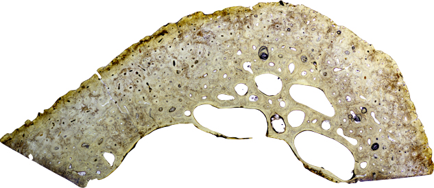
Image: Thin section of the humerus of a 17 year-old from St Gregory’s Cemetery.