A team of researchers are working with surgeons to install a unique AI-enabled combined Optical Coherent Tomography (OCT) and Raman spectroscopy system at Nottingham City Hospital to improve breast cancer diagnostics and guide surgery outcomes.
Researchers from the University of Nottingham have been awarded £1.8 million by the Medical Research Council to lead the project in collaboration with Kent and surgeons at the Nottingham University Hospitals NHS Trust to install the combined instrument at Nottingham City Hospital later this year.
Raman spectroscopy can detect molecular differences between normal tissue and cancer, but this requires hours to scan a whole specimen at high resolution. OCT can measure structural images of tissue at high speeds but lacks molecular specificity. AI algorithms specially developed for processing medical images will do a triage of the OCT images before they are processed further in the system.
The combined instrument maximises the speed and resolution of OCT and molecular specificity of Raman spectroscopy. This means that surgeons could analyse lumpectomy specimens during the time that a patient is in an operating theatre, allowing potential excision of breast cancer in a singular operation. Further surgery is often linked to poorer outcomes for patients, long recovery times and higher healthcare costs.
The project is being led by Professor Ioan Notingher from Nottingham’s School of Physics and Astronomy in collaboration with Professor Emad Rakha, Kent professors Adrian Podoleanu and Philippe de Wilde, and Mr Hazem Khout from Nottingham University Hospitals NHS Trust.
An initial OCT system designed by Kent has already been set up at Nottingham City Hospital, collecting images of breast lumpectomy specimens which were then processed by postgraduate using AI.
For the next phase of the project, this new OCT-Raman system is being assembled in Kent and will be set up at Nottingham City Hospital, for further research and evaluation to commence.
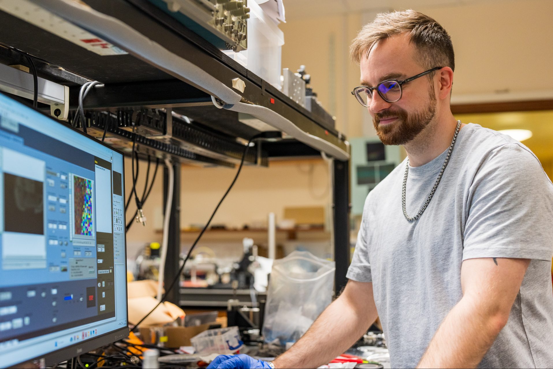
Postgraduate researcher, Dr. Radu Boitor, from Nottingham recently brought the Raman half of the system to Kent to be integrated with the OCT system and software assembled by postgraduate Kent researchers. Once the combined system is ready and tested in Kent, it will be transported to Nottingham. This requires demo testing before characterising the system on a variety of samples to perfect the software to make all processes more self-operating. A tunable laser, a galvo scanner and two large translation stages are under the OCT control designed in the Applied Optics Group. This leads the system to communicate with the Raman system, with a sensitive camera and proprietary software designed at Nottingham. The AI software sits in the middle of this.
Professor Podoleanu, is leading the assembly of the OCT part in Kent and said: ‘We have now reached a pivotal moment in this research project where we will see the combined system used in the hospital setting. By combining Raman with OCT there is hope that better diagnostics will be produced to guide breast surgery and outcomes.’
Professor Philippe De Wilde, supervising the AI algorithms, pointed out that AI algorithms for computer vision have great potential in new medical devices that can be produced in the UK, saying: ‘Our algorithms do not cost millions to train and run such as GPT, but run on a single processor. This is both economical and environmentally sustainable.’
Lead surgeon Mr Khout and Mrs Birgit Golden, a patient involved in the project, emphasised that the development of the OCT-Raman was ‘made possible thanks to the generosity of patients, who willingly participated, consenting to have tissue specimens scanned with this groundbreaking technology.’
This research is funded by the Medical Research Council.
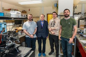
Part of the research team from left to right: Dr Rene Riha (Kent), Professor Adrian Podoleanu (Kent), Dr Marco Santopietro (Kent), Dr Radu Boitor (University of Nottingham).