
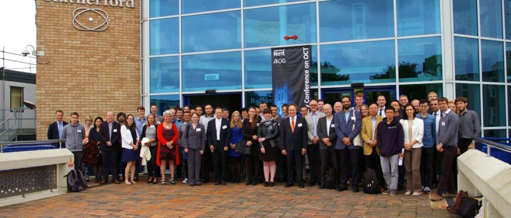
Over the last few days the Applied Optics Group hosted the 2nd Canterbury Conference on OCT in the maze of Rutherford College. The conference was chaired and organised by Prof Adrian Podoleanu as one of the activities of his UBAPHODESA European Industrial Doctorate training grant. I helped out here and there, including with reviewing submission, designing the programme and chairing one of the sessions. In total we had around 60 talks and posters, from participants from as far afield as South America, Japan, the USA and Australia.
The main website, including lots of photos, is at 2ccoct.aogkent.uk, and there is an article about the conference on the SPS blog.
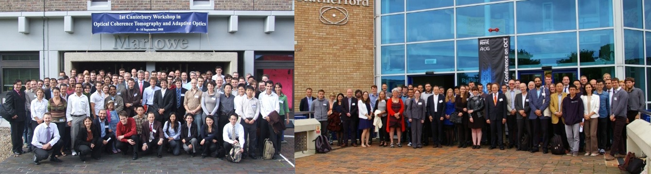
The meeting was particularly poignant for me, because I was also present for the 1st Canterbury Conference on OCT in 2008, in my previous life at Kent when I was studying for my PhD. I was less involved in the organisation back then, although I helped out with the website a little, but it was also my first ever conference presentation, on speckle reduction in OCT imaging of paintings.
This time around, the poster session included a poster by Miroslav Ďuriš, a student who visited us from Brno University for several months this year, and who worked with me on ultrathin endomicroscope probes. The abstract is below and proceedings will appear later in the year.
Towards an ultra-thin medical endoscope: Multimode fibre as a wide-field image transferring medium
Miroslav Ďuriš, Adrian Bradu, Adrian Podoleanu, Michael Hughes
Multimode optical fibres are attractive for biomedical and industrial applications such as endoscopes because of the small cross-section and imaging resolution they can provide in comparison to widely-used fibre bundles. However, the image is randomly scrambled by propagation through a multimode fibre. Even though the scrambling is unpredictable, it is deterministic, and therefore the scrambling can be reversed. To unscramble the image, we treat the multimode fibre as a linear, disordered scattering medium. To calibrate, we scan a focused beam of coherent light over thousands of different beam positions at the distal end and record complex fields at the proximal end of the fibre. This way, the input-output response of the system is determined, which then allows computational reconstruction of reflection-mode images. However, there remains the problem of illuminating the tissue via the fibre while avoiding back reflections from the proximal face. To avoid this drawback, we provide here the first preliminary confirmation that an image can be transferred through a 2×2 fibre coupler, with the sample at its distal port interrogated in reflection. Light is injected into one port for illumination and then collected from a second port for imaging.