The Applied Optics Group (AOG) has been at the forefront of fundamental research into Optical Coherence Tomography (OCT) for eye-imaging since 1996.
Optical Coherence Tomography for Biomedical Imaging
The University of Kent’s Applied Optics Group (AOG) is led by by Professor Adrian Podoleanu, who has over 27 years of experience in advancing Optical Coherence Tomography (OCT) technology for biomedical imaging. This highly accurate imaging technology is important for the diagnostic and treatment of various medical conditions ranging from glaucoma to skin cancer, benefitting millions of people across the world.
Researchers in the University of Kent’s AOG were the first to show en-face OCT images from the retina, now known as C-scans.
Building on this, they developed the world’s first combined Scanning Laser Ophthalmoscopes (SLO) and OCT instruments(OCT/SLO) for the diagnosis of eye diseases. The two-dozen patents protecting the original technology are still being maintained by key stakeholders that have successfully commercially exploited the technology.
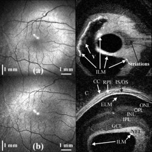
Pairs of confocal (left)/ultrahigh resolution C-scan images (right) of the macular region of the human retina in vivo: (a) the inner limiting membrane (ILM) and the vitreous (V) are visible; (b) the tilt in the C-scan sectionning plane with respect to the macula reveals retinal layers from a B-scan perspective: NFL nerve fiber layer, GCL ganglion cell layer, IPL inner plexiform layer, INL inner nuclear layer, OPL outer plexiform layer, ELM external limiting membrane, IS/OS junction between the inner and outer photoreceptors, RPE retinal pigment epithelium, CC choriocapillaris, C choroid. R. Cucu et al, •https://doi.org/10.1364/OL.31.001684
In collaborations with various academic and industrial partners, the Applied Optics Group continuously sought to further advance en-face technology ever since. Particularly noteworthy in this context is the collaboration between the University of Kent’s AOG, Richard Rosen at the New York Eye and Ear Infirmary (NYEEI) and Rishard Weitz at the Ophthalmic Inc Canada (OTI). The collaboration led to clinical advances of OCT/SLO and incorporation of other optical modalities, such as OCT/SLO with indocyanine green angiography (ICG), longer wavelength OCT/SLO, and ultra-high-resolution OCT/SLO.
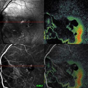
ICG/OCT/SLO sets of a patient with polypoidal choroidal neovascularization: Mid arterial-venous phase demonstrates leash of abnormal vessels with hyper-fluorescent bulbous ending. OCT image outlines the overlying serous elevation surrounding the vessels [R. Rosen, https://doi.org/10.1167/iovs.08-1855].
In collaboration with Optovue, Richard Rosen went on to develop and implement en-face views of vasculature of the retina, a technology now known as OCT angiography (OCTA). OCTA offers additional benefits to OCT as a non-invasive imaging technique that enables visualisation of vascular networks in the human retina, choroid, skin, etc. OCTA has been deemed a “landmark discovery” that “revolutionized the clinician’s access to non-invasive 3-dimensional vascular beds as a quantitative tool for management of diabetic retinopathy, age-related macular degeneration, and glaucoma”. OCTA also provided an impetus for new commercial developments, in particular the development of OCTA instrumentation.
AOG’s collaboration with DTU and Bispbjerg Hospital (Denmark), in turn, enabled the clinical application of combined OCT and laser technology, which has improved skin cancer diagnostics for thousands at Bispebjerg Hospital. OCT-laser technology is now being commercially exploited by companies, such as NKT.
The AOG has an ongoing programme for visual assessment with the Biomedical Research Centre at the UCL Institute of Ophthalmology – Moorfield Eye Hospital. In addition to being a research collaborator, the BRC has been involved as an associate partner in training PhD students on two Marie Curie Innovative Training Networks lead by Kent. One of these explores using tunable lasers for OCT (ongoing) and the other focuses on using OCT for broadband lasers commercialised by NKT.
OCT is mainly applied in ophthalmology but the AOG also has a programme on OCT for embryology in collaboration with Professor Darren Griffin (who developed a non-invasive imaging technology for embryo screening called Karyomapping with colleagues). Research ensuing from this programme has shown that time-lapse videos and Optical Coherence Tomography (OCT) can be used to measure developing embryo internal structure, . Being joined by Dr Alejandro Chavez-Badio this collaboration is now aiming to develop novel artificial intelligence (AI) strategies to improve assisted reproduction outcomes.
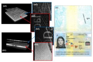
Further to this the AOG is evaluating the utility of OCT for forensic science and document inspection in collaboration with Foster and Freeman.